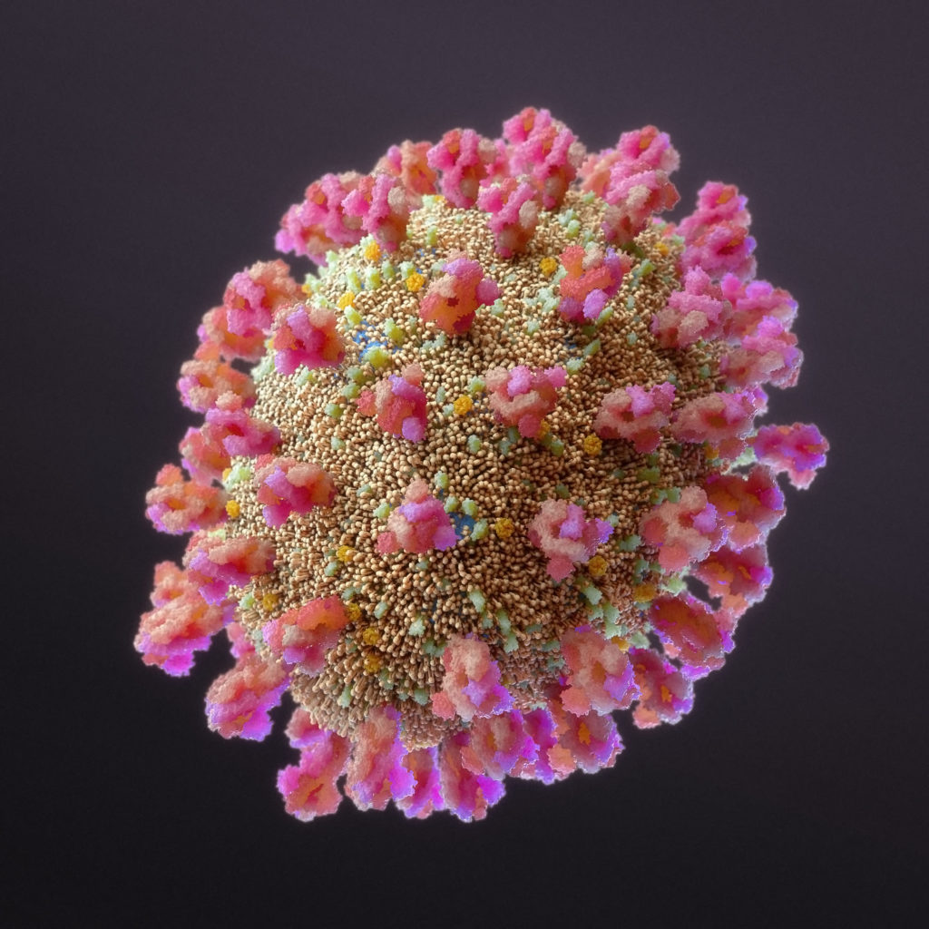
iSO-FORM LLC, Ames, IA, has created a SARS-CoV-2 3D image based on the most recent descriptions in scientific literature, scanning electron microscopy, and discussions in the medical visualization community. iSO-FORM…
Original size is 1024 × 1024 pixelsiSO-FORM LLC, Ames, IA, has created a SARS-CoV-2 3D image based on the most recent descriptions in scientific literature, scanning electron microscopy, and discussions in the medical visualization community. iSO-FORM LLC is a team of medical illustrators, software engineers, and artists whose focus is on bringing cutting edge science to targeted audiences using the latest techniques in visual effects, illustration, and biomedical visualization.
The image was created using VFX/Animation industry standard software. The molecular structures were created using ePMV and were assembled in Cinema4D. Most of the protein structures were acquired from electron microscopy or x-ray crystallography data. However, there are a few exceptions.
The E-protein is from the SARS-CoV virus, as information on this protein for the SARS- CoV-2 virus is not yet available.
The M-protein is computationally predicted and was acquired from DeepMind through predictive neural net processing.
The lipid bilayer was created in a VFX industry standard 3D software platform, Maxon Cinema4D, and was not derived from data, but is based on well-known lipid membrane quaternary structures.
SARS-CoV-2 is a spherical pleomorphic virus, which means it can be spherical, or often ellipsoidal in shape. Many coronavirus scanning electron micrographs show common ellipsoidal morphology, and thus, we have depicted it here as a variation on the more common spherical morphology.
Information on the structure and function SARS-CoV-2 is changing rapidly, and currently the organization of all proteins is an approximation based on current understanding. This model was not created by a simulation.